Nodular dermatitis is one of the most severe skin diseases that are found in cattle. Until recently, this disease was considered territorial, which is found on the African continent and nearby territories, but over the past few years, the infection has been able to spread widely into the depths of Eurasia. Currently, the disease poses a serious danger to livestock, but most farmers treat it with disdain. In this article we will take a closer look at what is nodular dermatitis, and also get acquainted with the main ways to combat it.
What is this disease
Nodular or nodular dermatitis is a complex infectious disease that occurs in cattle and other mammals. The cause of the development of dermatitis in livestock is the defeat of the body by a specific virus.  The infection is quite contagious, so it spreads instantly among animals, and also has serious consequences for the body of animals. Mortality of livestock from lesions of nodular dermatitis is from 4 to 95%.
Did you know? Cow's milk proteins are able to bind a lot of harmful toxins, which is why this product is a traditional free bonus for all workers in hazardous industries.
History of detection and dissemination
For the first time, people encountered this disease of livestock in 1929 in South Africa (North Rhodesia) and on the island of Madagascar. At this time, small point foci of infection appeared, which were perceived by many veterinarians as false urticaria.
Several decades later, in the mid-1940s, this pathology was identified by the British scientist Bakstrom as a list of individual diseases characterized by a high degree of infectiousness.
By the early 1950s, the disease began to massively meet in South Africa, in particular, in the territory of South Africa, Mozambique, Malawi and Namibia.
In 1960, outbreaks of infection reached the equatorial part of the continent, as well as North Africa, from which the infection spread to India and Romania in just a couple of decades.  In 2015, the illness reached the territory of the Russian Federation, large foci of infection were recorded in Chechnya, North Ossetia and Dagestan, and in 2017 in Tatarstan.
Today, nodular dermatitis is considered to be one of the most actively spreading infectious diseases of farm animals, and is also the main problem of industrial cattle breeding in Africa and nearby regions.
Pathogen, sources and routes of infection
The main cause of nodular dermatitis are pathogenic specific DNA viruses. Conventionally, they can be divided into three main groups: BLD, Allerton and Neethling. Often, livestock are affected by the Neethling group of viruses that are closely related to smallpox pathogens in goats, sheep and other artiodactyls.
Chlamydia, brucellosis, warts on the udder, EMCAR, blutang, leptospirosis, malignant catarrhal fever, anaplasmosis, parainfluenza-3, actinomycosis, abscess are also referred to infectious diseases of cattle.
This group of the virus is highly resistant to extreme conditions and viability, therefore, it can safely withstand up to 3 cycles of prolonged freezing outside the body's cells.
When mature, the Neethling virions are an independent structural unit of a rounded shape. It is characterized by a double shell, lateral inclusions, as well as a dense core with genetic material.
The development of the virus in the body occurs everywhere, but in most cases it affects the organs and adjacent systems responsible for the formation and active transport of various physiological secretions of the body (blood, semen, saliva, etc.).  The most susceptible to infection - cultural breeds of livestock, especially of European descent. Only one sick animal in 10-14 days can infect all livestock and cause a real epidemic.
The main reservoirs for the spread of infection are sick animals with a chronic or latent form of the disease, as well as active and passive carriers.
The virus spreads through the blood through blood-sucking insects. That is why the massive outbreaks of nodular dermatitis are recorded in hot countries, as well as the districts of mass reproduction of mosquitoes and mosquitoes.
Inside the body of insects, the virus successfully persists up to 1 month, which leads to almost uncontrolled transfer of the disease in any direction.
Also, the disease is actively spreading due to physiological secretions of animals. They are able to contaminate food, water and surrounding objects, and further reach an additional carrier - migratory birds.
The birds themselves are often not affected by a specific livestock virus, but rather successfully carry it with infected objects over considerable distances. The causative agents of nodular dermatitis do not differ in sexual or other preferences, therefore, they affect any cattle equally.  In addition, the development of the disease does not have a seasonal or regular nature, so it is almost impossible to control outbreaks of infections today.
Did you know? The tradition of Hinduism to worship a cow as a sacred animal has its roots in the Vedic culture dating from the I-II millennium BC. er
Incubation period and symptoms
The first symptoms of a lesion of the organism by the causative agent of nodular dermatitis occur in 3-30 days from the moment of infection, but often the incubation period of this disease is about 7-10 days.
The picture of the development of an infection depends on the general state of the organism, therefore the activity of the disease may closely depend on the ability of the immunity to resist the dangerous pathogen. The incubation period ends with a sharp increase in body temperature in infected animals up to +40 ° C.
Against the background of general heat in sick animals, the following symptoms are observed:
- loss of appetite;
- recurrent lacrimation;
- copious mucous discharge from the nose.
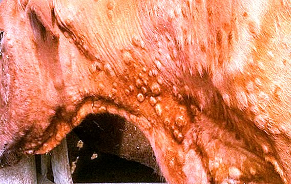
After 2 days after the temperature rises, the animals develop round or elongated nodules characteristic of the disease under the skin with a diameter of 0.5 to 7 cm and a height of about 0.5 cm.  The number of nodules depends on the degree of development of the disease, often the formations have a wide distribution - from a few dozen to several hundred. Sometimes single nodules may merge, in which case they form dense, convex spots.
After some time (1-2 days), the skin begins to separate along the edges of the nodules, and a small hollow appears along their center - this further leads to necrosis of the nodules and the appearance of characteristic putrefactive discharge.
2-3 weeks after the activation of the infection, the nodules are completely separated from the surface of the body, and in their place there is a dense scar, which eventually grows over with epidermis and hair. If the infection becomes more complicated, then ulcers appear on the site of the nodules.
During the period of active lactation nodules necessarily appear on the udder. In this case, this leads to a deterioration in the quality of milk. It becomes pinkish, thick, acquires an unpleasant odor and taste. After heating such milk turns into a thick gelatinous mass.
At the same time, there is inflammation of the lymph nodes in the cow, which is particularly pronounced in the subscapular region.
Important! If calves are affected by nodular dermatitis, the ailment is atypical. In this case, instead of the characteristic symptoms, the infection manifests itself with fever and recurrent diarrhea (without cutaneous manifestations).
With reduced immunity and the presence in the active phase of other ailments, the disease can occur in severe form.  In this case, the animal has:
- fever;
- severe loss of appetite and weight;
- labored breathing;
- gastrointestinal upset;
- nodules throughout the body, in the mucous membranes they appear as round ulcers and necrotic plaques of a grayish-yellow shade. Over time, they develop into abundant putrefactive lesions;
- ulcers and suppuration in the area of the eyes, this leads to erosion of the eyelids, as well as damage to the cornea and the eyeball;
- purulent mucus from mouth and nose.
Diagnosis of the disease
The diagnosis of cattle lesion with nodular dermatitis is made on the basis of:
- analysis of the mass infection - a clear sign of illness is a high infectiousness and extensiveness of distribution among the livestock;
- common clinical symptoms - a combination of heat, a sharp deterioration in the well-being of sick animals, as well as the manifestation of characteristic nodules on the skin;
- histological studies of nodules - in the cells of selected tissues, characteristic inclusion bodies are detected. They have the form of independent oval-shaped structures. The presence of pathological inclusions is confirmed due to cell staining with xanten dye (eosin);
- microbiological analysis - isolate viruses are isolated from the nodule tissue that infect sheep or calf embryos. They say about the specificity of the virus after the appearance of characteristic Taurus-inclusions in the tissues of embryos. Confirm the type of infection due to infection of susceptible animals (mice, sheep, goats, calves) and the manifestation of their characteristic signs of illness;
- differential analysis - using the data obtained in the course of the above studies, they differentiate the ailment from similar symptomatic diseases (urticaria, tuberculosis, strepto-trichosis, epizootic lymphangitis, demodicosis, smallpox, the effects of tick bites and other stinging insects, post-vaccination edema).

Pathological changes
Nodular dermatitis is an extremely dangerous disease, during the development of an infection, the virus causes severe changes in the body of animals.
Important! It is quite difficult to identify the ailment on its own at the first stages, often the first symptoms are blurred and do not have a clear picture of manifestation, therefore, when first suspicion of a lesion occurs with nodular dermatitis, you should contact a veterinarian as soon as possible.
As a result, the cattle observed:
- appearance of characteristic inclusions under the skin and in the area of muscle tissue, kidneys (under the capsule), lungs (in rare cases);
- swelling and swollen lymph nodes, often accompanied by watery tissues and an increase in the number of lymphocytes, eosinophils, plasma cells, neutrophils (with necrosis);
- recurrent hemorrhages in the visceral pleura, turbinate, capsule of the liver and spleen, in the scar tissue (after the destruction of the nodules);
- edema of the lungs and difficulty of the respiratory system;
- congestion and stasis in the glands, nasal passage;
- inflammation of the tissues of the rennet mucosa, often accompanied by ulcers in the bottom and pylorus;
- necrosis of the epidermis and the papillary layer of the dermis, along the edges of the damaged tissues there is a dense thickening of the dermis;
- perivascular infiltration of cells and blood clots in the veins are observed under the damaged tissue.
 In addition to the fallen animals, the following pathological changes can be found:
- signs of severe enteritis;
- hemorrhage in the mucous membrane of the colon and small intestine;
- lesions of the joints.
Treatment
At present, nodular dermatitis is safely treatable, for these purposes often use complex highly active drugs, characterized by powerful species-specific and multifunctional effects.
Among them, the most popular are the following drugs:
- "Biferon-B" - is a mixture of bovine interferon alpha-2 and gamma. The main active ingredients of the drug are in a stabilized form, therefore "Biferon-B" is distinguished not only by a powerful, but also by a long-lasting effect on the pathogenic virus. Enter the drug by intramuscular or subcutaneous injections. Animals weighing up to 100 kg of the drug is shown 1 time per day, with a calculation of 1 ml / 10 kg of weight. Cattle weighing over 100 kg of the drug is administered 1 time per day in a volume of 10-15 ml. The duration of therapy depends on the state of health of the animal, but often does not exceed 3-5 days;
- "Gentabiferon-B" - drug mixture consisting of gentamicin sulfate and bovine recombinant interferon alpha and gamma type. "Gentabiferon-B" is used for intramuscular and subcutaneous injections. Animals weighing up to 100 kg of the drug is administered 1 time per day with the calculation of 1 ml / 10 kg of weight. Cattle weighing over 100 kg of the drug is administered 1 time per day, in a volume of 15-20 ml. The duration of therapy is from 2 to 5 days;
- "Enrofloksavetferon-B" - The drug consists of antibiotic compounds from the group of fluoroquinolones enrofloxacin, as well as bovine recombinant alpha interferon. "Enrofloksavetferon-B" is administered by intramuscular injection with a calculation of 1 ml / 10 kg of weight, with an interval of 24 hours. The duration of therapy is from 3 to 5 days.
 In case of serious lesions of the skin, cattle skin is treated with antibiotic ointments 2-3 times a day, and synthomycin and zinc ointments, as well as Vishnevsky liniment, are best recommended for this purpose.
Important! Livestock products after highly active drug therapy are suitable for consumption not earlier than in 20 days.
To prevent the development of the effects of infection on the respiratory system and the intestines, in the treatment of livestock, an additional choice is used:
- "Nitoks-200" - use the tool as intramuscular injections with a calculation of 1 ml / 10 kg of animal weight. Enter "Nitoks-200" once, but if necessary, after 72 hours, repeat the injection;
- "Tetracycline" - used orally, every 12 hours for 5-7 days, with the calculation of 20 thousand. Units / kg of animal weight;
- "Oleandomycin" - use the drug intramuscularly, with a calculation of 20 mg / kg of animal weight 3 times a day. The duration of therapy is from 5 to 7 days.
Prevention and vaccination scheme
Despite the fact that animals that have been ill with nodular dermatitis develops resistant immunity to the ailment, high-quality and timely prevention is the main condition not only of successfully fighting infection, but also of preventing the general development of the disease in large areas.  The most effective preventive measures are:
- periodic inspection of animals;
- mandatory quarantine of sick individuals;
- a ban on the importation of livestock and animal products from potentially dangerous regions;
- active vector control of the disease.
Also, periodic vaccination of livestock will help protect against illness. Most often, complex vaccines or specific live preparations based on strains SP-143, Isiolo, and Kedong of lamb pox virus grown in an environment with lamb testis are used for these purposes.
Young animals are vaccinated for the first time at the age of 3 months, revaccination is carried out every 12 months. This allows you to create a massive and long-lasting immunity in the entire population.
Did you know? Vaccination as a method for combating dangerous infections was first applied in 1796 by the English doctor Edward Jenner to fight the smallpox virus.
Can a person get infected from sick animals?
Nodular dermatitis in cattle is absolutely harmless to humans, since today there has not been a single case of transmission from animals to humans.  However, it is recommended to contact with sick animals on the farm exclusively in protective gear, since a person can become a carrier of infected physiological fluids of sick cattle and, therefore, provoke an active spread of infection in large areas.
Nodular dermatitis is a complex infectious disease found in cattle quite widely. Despite the fact that large foci of this infection are often found in tropical and subtropical regions of Africa, each year the dangerous virus becomes closer to colder regions.
At the moment, this disease, unfortunately, remains not fully understood, so in order to avoid serious consequences for the health of animals, it is necessary to be vaccinated, otherwise nodular dermatitis can cause serious losses.